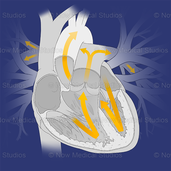
Heart cross-section systole. Stock medical illustration
A medical illustration of the cross-sectional and internal anatomy of the heart. Here you can see arrows indicating the flow of blood during systole (the phase of the heartbeat when the heart muscles contracts and eject the blood to the aorta and pulmonary trunk).
Here you can see anatomical landmarks of this major cardiovascular organ; such as the ascending aorta, brachiocephalic trunk, left common carotid artery, left subclavian artery, left pulmonary artery, left and right pulmonary veins, left and right atrium, superior and inferior vena cava, aortic valve, tricuspid valve, mitral valve, chordae tendineae, papillary muscle, intraventricular septum, fossa ovalis, epicardium, myocardium, endocardium, and more.
These illustrations were created on a transparent background, giving you full flexibility to use them with your brand colours.
Please reach out to us at hello@nowmedicalstudios.com if you have other licensing requirements not listed in our options below.
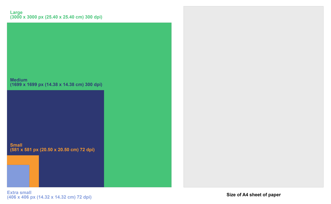
Image sizing guidelines
Below is an example of what image sizes look like in comparison to a standard A4 sheet of paper.
Further details about the image
Credit: Now Medical Studios
Licence type: Rights-Managed
Location: Scotland
Release info: Property released
Serial Number: 0019


